Hip disease


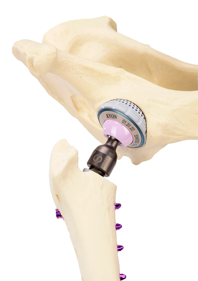
Total Hip Replacement is now accepted as best way to manage long term hip disease in dogs and cats. It is a technical and difficult procedure. There are few surgeons experienced enough to offer a high chance of success. Our hip surgeon, Dr Stuart Cooke has performed over 500 Total Hip Replacements and is the lead UK tutor for Kyon, one of the world’s foremost manufacturers of hip implants. Our team offers your pets the best chance of a good outcome from their Total Hip Replacement.


The most common condition requiring THR is hip dysplasia. Surgery can also be indicated with femoral head or neck fractures, pelvic (acetabular) fractures, traumatic hip luxation (dislocation) and Leg Calfe Perthes Disease. Hip disease can remain hidden for a very long time. It is rare that a pet is snappy or makes noises, they suffer in silence. You need to look out for stiffness on getting up, seeming slower or older, reluctance to climb stairs, hesitating before hopping onto the bed or jumping into the car.


The first step in a pet’s investigation is a consultation, this is then followed by investigative tests such as xrays or CT scanning to assess the hips and the other joints such as the lumbosacral junction. The findings are discussed at a discharge consultation and a plan is made, surgical or non-surgical.

What are potential treatment options?


When hip dysplasia, hip injury or severe arthritis are treated properly by hip replacement, you can expect your dog or cat to have a complete, functional recovery. Your dog may even return to agility and sporting activities.


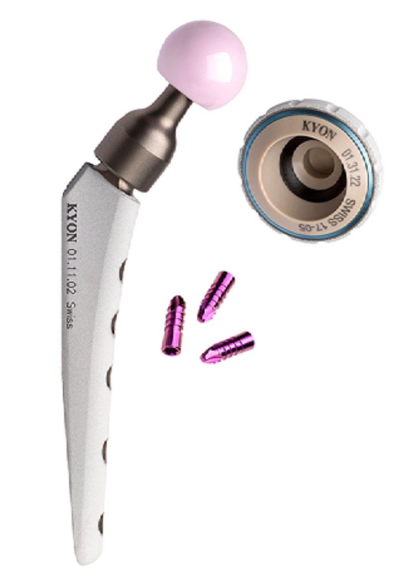
Our objective is to provide patients, often less than 1 year old, with a device that will last a lifetime. The implants use the best materials available for medical implants, are manufactured from Titanium, PEEK and Ceramic. Stems and cups are coated with hydroxyapatite, a surface treatment proven to promote bone integration. Proprietary locking screws secure the stem, give the patient immediate use of their leg after surgery and reduce common complications associated with other canine THR systems. Patented geometry of the cup inlay reduces stress and wear by several fold.

Recovery after hip replacement surgery


Your cat or dog will be able to walk out of the hospital the following day after surgery, but he or she needs rest for their hip to heal. Most dogs feel so much better shortly after surgery that they want to play and run, but you will need to keep your dog quiet with restricted exercise for two months to ensure longterm success. This means crate rest, leash-only walks, and preventing play or rough housing with other dogs or family members. If needed, a mild sedative can be prescribed. Complications are rare, typically occur within the first two months after surgery and are significantly reduced when you follow all post-surgical recommendations. If a complication does occur, the Zurich Cementless hip replacement system has proven to be extremely versatile in resolving even the most complicated revisions.


How much does it cost?


The initial consultation, surgery and standard aftercare typically costs around £9000 (£8000-£10000 depending on the patient and plan).

What is canine hip dysplasia?


Hip dysplasia in dogs is a genetic disease that results in an abnormal formation of the hip joint(s). The hip joint functions as a ball and socket that should work together by sliding smoothly. When the ball and socket do not fit together or develop properly, the joint rubs and grinds against each other and results in the deterioration of the joint. This lifelong condition not only deteriorates the joint but also diminishes your dog’s active life.


What causes hip dysplasia?


Various factors contribute to the development of hip dysplasia, but the leading factor is genetics. Hip dysplasia can be present in the canine genoma even if the parents are not affected. The severity of the disease can be influenced by the environment and worsened by excessive body weight and activity. Many breeds can be affected, like Labrador, Golden Retriever, Bulldog, Germans Shepherd, Rottweiler, Border Collie and many other including small breeds and mongrels. In the United States 20-30% of dogs are impacted by canine hip dysplasia.


What should I look out for?


Severe hip dysplasia can easily go unnoticed and undetected by you. Your dog could be hiding the pain for years, until the discomfort and instability gets so bad that he or she is having difficulty maintaining normal activities. Symptoms include: • Decreased activity • Abnormal gait shifting the body weight on the fore limb by curving the spine and lowering the head while walking • Difficulty with activities: raising from sitting, climbing stairs, jumping and running • Lameness in the hind end when one hip gets worse • Walking with short steps and running like a rabbit • Enlargement of shoulder muscles (compensation) • Change in personality, a once happy and excited dog becomes lethargic and unmotivated Typically, dogs react immediately to acute and unexpected pain, but dogs with chronic hip pain mask their discomfort until it becomes unbearable. Before then, it may seem as they act older than they are. X-ray imaging will confirm the severity of osteoarthritis.

THR – What is total hip replacement for dogs and cats?

THR – How to care for your dog after total hip replacement surgery?

Total Hip Replacement – 6th Generation

Total Hip Replacement – Dual Mobility

Cranial Cruciate Ligament Degeneration and Rupture


Cranial (anterior) cruciate ligament damage is the most common condition in veterinary orthopaedics. It can cause anything from insidious arthritis to sudden & complete joint instability and loss of the use of the leg. This is surgical problem; surgery is the route to a better future. If a dog or cat is lame in the hindlegs this is the condition we need to diagnose or exclude first. Various techniques are available to manage this problem. We carry out one of four techniques: Tibial Plateau Levelling Osteotomy (TPLO), Cranial Closing Wedge Osteotomy (CCWO), Tibial Tuberosity Advancement (TTA) or Extracapsular repair (ECR or Lateral Suture).

We want concerned pet owners to feel confident our patients are being looked after by a team that cares for pets and people, in a location that is correctly equipped, designed and maintained, to give every pet the best chance of a happy and healthy life after the surgery. We diagnose CrCL rupture using a combination of examination and xrays under sedation. This is a day patient procedure, your animal companion will be home with you by the evening. The surgeon will discuss the diagnosis and surgical plan during the discharge consultation.


Surgery aims to stabilise the knee so that it no longer requires a natural Cranial Cruciate Ligament. This is achieved through cuts in the bone and metal implants (TPLO, TTA, CCWO) or placing a prosthetic ligament outside of the joint, underneath the muscles (ECR). Surgery involves an overnight stay to allow reliable pain relief to keep our patients comfortable. We get them home to you the day after the operation, along with a rest and exercise plan.


Most pets are as comfortable as they were before the surgery within 2 weeks, and back to running around 10 weeks later. Some pets need long term pain killers to manage the arthritis they had before the surgery but most of our patients go on to live a long and fulfilled life for many years without pain killers.


Without surgery, patients suffering CrCL damage invariably go on to develop arthritis, pain in the knee and a reduction in the quality of their welfare. If a patient is suffering severe debilitating illness or conditions in multiple limbs and/or the spine, surgery may not be appropriate; we will discuss and determine the best plan for each individual pet.


How much does it cost?


The initial consultations, X-rays, surgery and standard aftercare cost from £3000-6000 depending on the surgery required, patient and overall plan.

Elbow Dysplasia and Pain


Elbow dysplasia and medial compartment disease are the most common causes of forelimb lameness in dogs. The elbow is a complex joint and there is no one size fits all approach to the problems. We take a holistic view of your pet’s elbow problem and manage each case individually. We determine the best approach to each patient based on examinations, progress, the results of tests and responses to therapy.

CT scanning allows detailed examination of an elbow. Typical elbow problems include Humeral intercondylar fissure (HIF), Fragmented Medial Coronoid Process of the Ulna (FCP), humeral OCD, United Anconeal Process of the Ulna (UAP) and joint incongruency (poor fit between the bones). Ongoing lameness in a dog with CT results confirming elbow pathology (disease) often leads onto elbow arthroscopy. Arthroscopy is the examination of the inside of the joint using a very small (1.9-2.7mm wide) telescope. Treatments can also be delivered at the same time: removal of damaged tissue (debridement) and cutting out of fissured bone (coronoidectomy). If arthroscopy does not result in a long term improvement there are further non-surgical and surgical options.


Surgical: PAUL to shift the weight away from the painful elbow tissues; CUE to replace damaged cartilage with a  prosthesis. One day we will have the option of using a Total Elbow Replacement but there is not yet any implant good enough for us to offer this to our patients.


Non-Surgical: Injections to the joint (intra-articular therapy) using either corticosteroids, hydrogels, local anaesthesia, Platelet Rich Plasma (ACP/PRP) or Stem Cells; multi-modal pain relief & anti-inflammatories, diets, exercise and physiotherapy.

HIF screening


Certain breeds of dog are prone to incomplete fusion of the humerus, a humeral intercondylar fissure (HIF), this can lead to fractures of the humerus:

  • Springer Spaniel
  • Cocker Spaniel
  • Staffordshire Bull Terrier
  • French Bulldog
  • Golden Retriever
  • Labrador
  • Basset Hound
  • Newfoundland
  • German Shepherd
  • Rottweiler

We advise CT scanning the elbows of these breeds at or after 18 months of age. If a HIF is diagnosed we advise placement of a transcondylar screw to reduce the risk of complex fractures of the humerus, which can follow even innocuous forelimb injuries.

Patella Luxation


The knee joint (stifle) consists of the patella (knee cap), femur (thigh bone) and tibia (shin bone). The patella should sit in the groove at the end of the femur, the trochlear sulcus. In some cats and dogs the patella can slip out of position, this dislocation is a problem called patella luxation. Patella luxation is graded from 1 to 4. The grades we most commonly operate on are Grades 2 and 3.

Grade 1: Patella sits in the normal position but we are able to briefly dislocate it on examination.

Grade 2: Patella spontaneously slips in and out on examination, returning to a normal position.

Grade 3: Patella sits out of place but it can be moved back into the normal position during examination before it dislocated again.

Grade 4: Patella is permanently dislocated and cannot be moved back into its normal position.


The severity of each pet’s problem depends on their size and shape. Small dogs and cats can tolerate this better than heavier muscled pets. We base our recommendations on each patient, not only the grade of the luxation. We only advise surgery when we believe a pet will benefit from it. Assessment does not invariably lead to surgery. The patella is a 3-dimensional joint and each patient has a unique combination of issues, anywhere from the femoral neck, near the hip, to the tibia. We use X-ray or CT imaging to plan the surgery. We use a range of techniques to manage this problem and will explain the surgical plan based on your pet’s own unique shape:


  • Tibial Tuberosity Transposition, moving the attachment of the patella so that it wants to glide in the correct groove.
  • Sulcoplasty, deepening the groove the patella rides within.
  • Fascial Release, loosening of the attachments that pull the patella out of position.
  • Imbrication, tightening of the tissue to encourage the patella to remain in position.
  • Femoral osteotomy, removing a wedge of bone from the femur to straighten and re-align the knee joint, then stabilising the cut femur with a bone plate and screws.
  • Patella Groove Replacement, a titanium prosthetic replacement of the femoral sulcus. This is very useful in dogs that have suffered trochlear injury, total loss of cartilage or a condition called patella alta.

Pets remain in the hospital overnight after their operation for pain relief and go home the next day. Owners need to rest their pets for several weeks after the operation to give their pets the best chance of healing properly. We prescribe pain killers to keep our patients comfortable and we take x-rays 6-8 weeks after the operation to plan our patients’ return to free exercise. As long as a cat or dog is not suffering marked arthritis before the operation we expect our patients to be able to move better, without pain killers, for many years.

Brachycephalic Obstructive Airway Syndrome (BOAS)


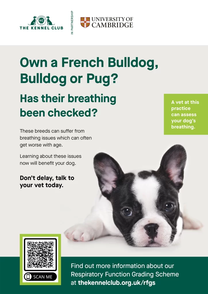
Short nosed breeds of dog are now very popular (Brachycephalic dogs). French Bulldogs, Pugs, Boston Terriers, English Bulldogs and many more are busy, bustling characters, tons of fun and adorable. Breeding for a compressed shape to the head means that our pets’ breathing is impaired. Even small reductions to airway width make a huge difference: try pinching your nose.


The narrow nostrils are visible to the naked eye but other deeper issues make the problem worse: excessive and dense tissue in the nasal cavity, an overlong and thickened soft palate, enlarged tonsils, an oversized tongue, collapse of the larynx and a narrow trachea. We often perform CT scanning of the head to confirm what deeper changes are present and help predict what issues may develop in the future.

Thankfully, we can improve brachycephalic dogs’ quality of life and make every breath easier for them. Surgical correction of their airways is a great gift. After surgery, dogs will be much less at risk of overheating in warm weather, less prone to regurgitation and gagging when running around having fun, they will be fitter and less restricted. The common technique is to widen the nostrils (nares) and shorten the soft palate. Sometimes we also need to remove the tonsils and some loose pockets of tissue from the larynx (laryngeal saccules). We use electro-surgery to minimise the risk of post-operative bleeding or swelling and aim to have your pet home with you the day after the operation. Most dogs make an uneventful recovery and lots of owners report that they too sleep better because their dogs snore so much less!


Alice Webb-Wilson our head vet at Chiswick is an RCVS Advanced Veterinary Practitioner in Small Animal Surgery and also an accredited respiratory function grading assessor with The Kennel Club.